Anatomy is the backbone of clinical practice, enabling doctors to drive through the body’s complex anatomy with accuracy. Two fundamentals of the lower limb’s saphenous vein and the neck triangles are not only theoretical ideas but also hands-on books in surgery, diagnostics, and treatment planning.
The Longest Saphenous Vein in the Human Body
The saphenous vein is a significant superficial vein of the lower limb and is the longest body vein. Since it lies just under the skin, it is visible in thin people and can conveniently be used for certain medical interventions. The saphenous vein comes in two broad forms:
- Great Saphenous Vein (GSV)
- Origin: It arises from the medial margin of the dorsal venous arch of the foot.
Course:
- Passes in front of medial malleolus (bony prominence on medial ankle).
- Rises along the medial aspect of thigh and leg.
- Passes through a saphenous opening in fascia lata.
- It got emptied into the femoral vein at the saphenofemoral junction of the groin.
- Tributaries: It will Arise from a group of superficial veins, such as superficial epigastric, superficial circumflex iliac, and superficial external pudendal veins.
Clinical Significance:
- Most commonly used in coronary artery bypass grafting (CABG) because of its ideal length and diameter.
- Usually resected in peripheral vascular surgery.
- Susceptible to varicose veins because of incompetent valves.
- Small Saphenous Vein (SSV)
- Origin: It Arises from the lateral tip of the dorsal venous arch of the foot.
Course:
- Run behind the lateral malleolus.
- Runs upwards along the posterior midline of the leg.
- Terminates in the popliteal vein within the popliteal fossa (behind the knee).
Clinical Importance:
- Easily employed for calf deep vein thrombosis (DVT).
- Sometimes employed as a graft for the smaller veins.
Roles of the Saphenous Vein
- Permits oxygen-poor venous blood to flow from superficial lower limb tissues back into deeper veins.
- Employed as a site for intravenous cannulation or venous cutdown during an emergency scenario.
- Serves as a source of surgical graft.
Normal Abnormalities:
- Varicose veins: It got Enlarged and coiled veins due to incompetent valves.
- Thrombophlebitis: Inflammation caused by clotting.
- Venous insufficiency: Inadequate blood return leading to swelling, pain, and changes in the skin.
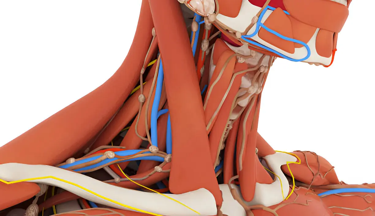
Triangles of the Neck – Anatomical Road Maps
The neck harbors is major neurovascular elements, glands, and lymph nodes. Anatomical exploration and surgical approach are aided by dividing the lateral triangles of the neck over surface landmarks constituted by the SCM, trapezius muscle, and inferior mandibular border. Surgeons, radiologists, and clinicians will receive the assistance in the identification of particular structures from these triangles.
- Anterior Triangle
Subdivisions:
- Submental Triangle – Carries submental lymph nodes and tributaries of anterior jugular vein.
- Digastric Triangle – It will Contain the submandibular salivary gland, facial artery, and vein.
- Carotid Triangle – Carries bifurcation of common carotid artery into internal and external carotid arteries, internal jugular vein, vagus nerve, and hypoglossal nerve.
- Muscular Triangle – It have infrahyoid muscles, thyroid gland, and parathyroid glands.
Clinical Relevance:
- Surgeons will perform the carotid endarterectomy with the help of carotid triangle to excise arterial plaques.
- Submandibular triangle under edema may present with salivary gland infection or tumor.
- Muscular triangle is checked for thyroid enlargement (goiter).
- Posterior Triangle
Boundaries:
- Anteriorly: Posterior border of SCM.
- Posteriorly: Anterior border of trapezius muscle.
- Inferiorly: Middle third of clavicle.
Subdivisions:
- Occipital Triangle – Transmits spinal accessory nerve, branches of cervical plexus, and vessels of transverse cervical.
- Supraclavicular (Subclavian) Triangle – Has subclavian artery, subclavian vein, and supraclavicular lymph nodes.
Clinical Significance:
- The main Region for central venous has the access of the subclavian vein.
- Palpation of the supraclavicular lymph node can identify abdominal or thoracic malignancies.
- Infections or the metastasis result in swellings in the neck, which are initially found here.
Also Read-Innovative Test Generation Techniques for Modern Applications